
chromatographic separation assignment exploring the techniques to separate mixed liquids
Question
Task: how chromatographic separation assignment techniques be implemented towards separation of mixed liquids?
Answer
Introduction:
This chromatographic separation assignmentexplores methods to separate mixed liquids. The chromatography is defined as the study of separation of molecules mainly depends on the differences in their structure, elements and composition. The chromatography includes the sample which require to dissolve in the mobile phase i.e. Liquid, gas or supercritical fluid. However, the mobile phase is require to forcethrough theim-mobile, immiscible stationary phase. The phases are elected such a way the elements or component of the sample have distinct solubility in each phase. The component considered on this chromatographic separation assignment is a considerable solution in the stationary phase will take considerable time in order to travel through that and then the component which is not effective solution in the stationary phase but effective solution in mobile phase. The outcome of that difference in the mobilities, the sample element will become separated from each other as they travel using stationary phase (Omata, T. and Murata, N., 1983).
In this chromatographic separation assignment experiment, the chromatographic separation can be conduct by using different support that includes im-mobilization slice over the glass plates, volatile gases, paper and liquid that may incorporate hydrophilic, insoluble molecules. The column chromatography is the alternative iso-lation and purification approach uses considerable by organic chemists to find the pure sample of chemical from the natural resources or from the specific reaction sequences.
In case, there is compound separation in the column chromatography are colored than the specific procedure of the separation can be effectively monitor through visual approach. in fact, this is more general that the compound to be separate from the column chromatography are color-less. For that, there are several application for evaluation and inspection the separation progress has been planned. This is the simplest application of those includes the collectionof considerable small fraction of the eluent in labelled tubes and analysis of the components of particular fraction through the technique of thin layer chromatography (Sato, N. and Murata, N., 1978).
Aim:
To perform the chromatographic separation assignment laboratory experiment using chromatographic techniques through qualitative analysis and thin-layer chromatography, and column chromatography elemental procedure.
Objectives:
To perform quantitative analysis
To perform laboratory experiment using thin-layer chromatography and column chromatography.
Conduct experiment using mixture separation method.
Methods:
The following is provides the chromatographic separation assignmentexperimental procedure of chromatography of the spinach pigments.
Arrange the glass wool at the bottom section of the column.
Place approximate 10mL 7:3 petroleum ether and acetone solution in the column.
Approximate weight with 20 gram of silica gel, 40 in a 50 mL erlen-meyer flask, and added sufficient amount of petroleum ether acetone solution whereas, the mixture of the slurry with the glass rod.
Arrange the slurry in the column through pipette unceasingly, than arrange the flask under the column and allows to excess solvent to the drain.
Collect the addition solvent that can be re-used to place the column.
Sample preparation: spinach:
Place the spinach into the mortar, added 22 mL acetone, approximate 3 mL petroleum ether and spatula tip of calcium carbonateKhalyfa.et.al.(1992).
Grill the leave until the complete pieces are collect into considerable small segment, that to be seen and further filter the pigment removal, if essential.
Place the filtration into the separatory funnel and added approximate 20 ml petroleum ether + 20 ml with 10% NaCl solution.
Mixed well the funnel and drain the lower layer into the beaker.
Completely wash the upper layer two time with 5 mL distilled water.
Place the extract into the 50 ml flask and add the spatula tip of the sodium sultfate.
chromatographic separation assignmentexperiment column chromatography:
By using the pipette, added approximate 10 mL leaf as removing from the top of the column.
Open the bottom outlet of the column, and allows the elute passes through the column effectively. As eluent passes down the column, the sample is prepared.
If the yellow bend reach the bottom segment of the column, that is require to collect the layer test tube.
Change the test tube as observing eluent varies in color. This way, the separation is obtained.
If require, the evaporate the additional solvent in the sample, and close the sample test tube using parafilm and store.
Observations;
By Bruce Banner:
TLC notes:
Distance travelled.
A-Aspirin: 4.4 cm
P-Paracetamol: 3.2 cm
C- Caffeine: 1.4 cm
M1: mixture 1 : 4.7 cm
M2: mixture 2 : 2.75 cm
M3: mixture 3 : 1.4 cm
S: solvent front: 6.65 cm
R_f (A)=A/S
Remember for my RF calculations:
Two decimal places
No units.
Results:
Below is an image of Bruce’s TLC plate as it appeared under the UV light.
The notes in his lab manual were taken by measuring the distance each spot travelled following the TLC procedure.
Consider if the TLC worked as you may have expected.
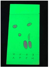
Image of Bruce’s column, following separation of mixtures.
Use the practical manual notes to see if you can identify any of the coloured bands observed in the picture. Johnstonet.al.(2013).
Are all the expected compounds identifiable
Consider if there are any improvements that could be made in future.

Sara Sidle’s data and pictures taken in the laboratory
Sara Sidle Laboratory tests:
TLC notes:
Distance travelled by solvent front = 6 cm
Distance travelled by each substance
C- Caffeine: 1.6 cm
A-Aspirin: 4.3 cm
P-Paracetamol: 2.8 cm
Unknown substance:
First spot : 1.7 cm
Second spot : 2.9 cm
Third spot : 4.5 cm
Ranout time require to check procedure guide for RF calculation.
Below is an image of Sara’s TLC plate as it appeared under the UV light.
The notes in her lab manual were taken by measuring the distance each spot travelled following the TLC procedure.
Consider if the TLC worked as you may have expected.
Perhaps there are things Sara could have improved that could be considered in the discussion of results.
Image of Sara’s column, following separation of mixtures.
Use the practical manual notes to see if you can identify any of the coloured bands observed in the picture.
Are all the expected compounds identifiable
Consider if there are any improvements that could be made in future.
Discussion:
From this chromatographic separation assignmentexperiment, the pigments from the spinach were collected through the column chromatography and extraction analysed through the thin layer chromatography. There are mainly two major pigments in the chloroplast i.e. chlorophylls and carotenoids. It observes the differences in the polarity of the spinach pigments that allows the isolation and separation of the individual pigment through column chromatography and separation was observes through color difference in the pigments. It observes that the green band collection for chlorophyll and yellow band collection for carotenoid.
Using column chromatography, the pigment were collected in order from the least polar pigment to the most polar pigment. The pigments separation was evaluation through visualization on the TLC plate and calculation and compare the retention factor value. For this case, the visualization and calculation of Rf suggest that the existence of chlorophyll “a” and “b” and pheophytin “a” in the extraction sample and presence of the chlorophyll pigments in the green band of the column chromatography (Jubert, C. and Bailey, G., 2007).
For this case, the column chromatography includes the partition of compound though the difference between the mobile and stationary phases. In that the stationary phase function as the adsorbents, anhydrous sodium sulfate and alumina in that chromatographic separation assignment experiment, through which the mobile phase is forwarded.
Further, the thin layer chromatography is the specific method of the separation and observes the mixture of two or more compounds using the allocation of the mixture between the mobile and stationary phases. From this experiment, it observes that the solid liquid procedure applied, in which the stationary phases was the solid whereas the mobile phase was the liquid. The solid phase was the plastic plate covered through the adsorbent, specifically, silica gel.
Lastly, it observes that the top-most orange band of the pigments in the separation resembles to carotene. However, the yellowish band appearing below that suggest the xanthophylls. Lastly, the third form would be dark green band demonstrate the chlorophyll “a”. The least yellowish green band is behave as chlorophyll “b”.
It is recommend to use spinach leave must be fresh and green. It should observes that the loading spot should be 2-3 cm away from the tip of the notch area.
Conclusion:
From the chromatographic separation assignmentexperiment, it observes that the spinach extract contains more than one photosynthetic pigment which has different absorption rate. From the experiment, it observes that the spinach extract contains the different photosynthetic pigments which observes the noticeable amount of absorbed the solvent at the different length as demonstrate in above experimental results.
The experimental results and inference of the procedure is which the obtained outcome are effective, accurate but they may not seem to have considerable difference of the equal experiment done in previous. Overall, the chlorophyll “a” to chlorophyll “b ratio should be 3 :1 and varies accordingly. In general, there is much further from 2.6 :1, that is the specific ratio of the two chlorophylls observes used for this chromatographic separation assignment expereiment.
References:
Jubert, C. and Bailey, G., 2007. Isolation of chlorophylls a and b from spinach by counter-current chromatography. Journal of Chromatography A, chromatographic separation assignment1140(1-2), pp.95-100.
Johnston, A., Scaggs, J., Mallory, C., Haskett, A., Warner, D., Brown, E., Hammond, K., McCormick, M.M. and McDougal, O.M., 2013. A green approach to separate spinach pigments by column chromatography. Journal of Chemical Education, 90(6), pp.796-798.
Khalyfa, A., Kermasha, S. and Alli, I., 1992. Extraction, purification, and characterization of chlorophylls from spinach leaves. Journal of Agricultural and Food Chemistry,chromatographic separation assignment 40(2), pp.215-220.
Omata, T. and Murata, N., 1983. Preparation of chlorophyll a, chlorophyll b and bacteriochlorophyll a by column chromatography with DEAE-Sepharose CL-6B and Sepharose CL-6B. Plant and cell physiology, 24(6), pp.1093-1100.
Sato, N. and Murata, N., 1978. Preparation of chlorophyll a, chlorophyll b and bacteriochlorophyll a by means of column chromatography with diethylaminoethylcellulose. Biochimica et Biophysica Acta (BBA)-Bioenergetics, chromatographic separation assignment501(1), pp.103-111.