
Biology Assignment: Questions and Answers
Question
Task:
Question 1: Immune System (1.5 Marks)
Which type of immunity is expressed in the following scenarios Choose between:
- Non-specific (first line defence)
- Non-specific (second line defence)
- Specific (antibody-mediated)
- Specific (cell-mediated immunity)
|
Scenario |
Type of immunity |
|
Coughing or sneezing. |
|
|
The hypothalamus raises the body temperature above the normal range, producing fever. |
|
|
Plasma cells respond to chicken pox virus |
|
1. Question 2: Immune System )
Choose TWO of the below scenarios and highlight these rows bold. State which general property of adaptive immunity the scenario illustrates in the second column. Select ONE phrase (from the list below) that BEST describes what is happening at a cellular level to result in this property of adaptive immunity. Write the letter corresponding to this phrase in the third column. An example is included. (0.5 mark each – total 2 marks)
|
Scenario |
General property of immunity |
Explanation |
|
Even though your body contains a relatively small number of lymphocytes, your immune system can mount a response against almost any antigen it encounters. |
Versatility |
A |
|
Generally, after an initial infection, subsequent immune responses to that antigen are faster, stronger and more sustained. |
|
|
|
You can suffer from ‘the flu’ multiple times throughout your lifetime. |
|
|
|
Your immune system can distinguish between antigens on your own cells, and those of an invading pathogen. |
|
|
Possible phrases are listed below. Not all phrases will need to be used and only ONE phrase is required per row. If you record more than one, you will not be awarded any marks for your explanation.
|
A |
There are millions of different lymphocyte populations, each of which is sensitive to a different antigen. Lymphocytes proliferate when activated by their specific antigen. |
|
B |
Each T or B cell has receptors that respond to only one antigen and ignores all others. |
|
C |
Activated lymphocytes produce two groups of cells: one group that attacks the invader immediately, and another that remains inactive unless it is exposed to the same antigen at a later date. |
|
D |
Each T and B cell can recognise many antigens and respond to a wide variety of possible threats. |
|
E |
T cells are versatile because they produce copious quantities of antibodies that can respond to a wide variety of threats. |
|
F |
Phagocytes have a reduced ability to destroy pathogens during subsequent infections. |
|
G |
The immune response ignores self-antigens and targets non-self antigens. |
|
H |
B cells differentiate into T cells to create a long-lived immune response. |
Question 3: Bones )
Describe the role of osteoblasts and osteoclasts in the regulation of blood calcium ion concentration. You must include the role of the relevant hormones for full marks.
Question 4: Muscles
Define atrophy and use an example to explain how this term relates to skeletal muscle tissue. (3 marks)
Question 5: Endocrine System
Think about how the renin-angiotensin-aldosterone system acts to maintain normal blood pressure. Susan has sustained damage to her adrenal glands. Consequently, her circulating levels of aldosterone are abnormally low. What do you expect the lack of aldosterone to do to Susan’s blood pressure Explain your answer.
Question 6: Endocrine System (2 Marks) And Urinary System
Tasma is enjoying a night out at the pub with her friends. It is well established that alcohol inhibits the secretion of ADH. Explain how this will affect her urine output. (3 marks)
Question 7: Genetics
Duchenne muscular dystrophy (DMD) is recessive sex (or X)-linked disorder, which causes muscle degeneration and premature death. A couple is pregnant with a boy and neither has DMD. The father’s family has no history of DMD, but the mother is unsure of her genetic family history as she was adopted. The mother decides to take advantage of some of the new genomic screening tests and learns information about a large number of her genes. She finds that she is a carrier of the DMD-affected allele. (3.5 marks total)
A. What is the mother’s genotype Explain your reasoning.
B. What is the father’s genotype Explain your reasoning.
C. Explain why DMD is more common in males than females
Question 8: Respiratory
Describe the changes in volume and pressure inside the chest cavity during a normal, quiet exhalation.
A. Explain why these changes are occurring, and the effect upon airflow.
B. Is quiet exhalation an active process Briefly explain your answer.
Question 9: Respiratory System
A. Complete the below table of partial pressures in external respiration.
|
|
pO2 (mmHg) |
pCO2 (mmHg) |
|
Alveoli |
|
|
|
Capillary |
|
|
B. Use to the figures above to explain how differences in partial pressures drive the direction of gas movement between the blood and alveoli.
Question 10: Cardiovascular System
A person with type B blood has been involved in a car accident and excessive bleeding necessitates a blood transfusion. Due to an error by a careless laboratory technician, the person is given type A blood. Explain what will happen. (4 marks)
Question 11 : Cardiovascular
To maintain adequate circulation, the average resting heart rate of a healthy non-athletic person is between 60-90 beats/minute. However, a trained athlete’s heart rate is usually about 40 beats/min (sometimes as low as 30 bpm!). Despite this, adequate circulation is maintained in these athletes. Using the terms cardiac output and stroke volume, explain how this is possible. (3 marks)
Question 12: Cardiovascular
A. The Wigger’s diagram (below) represents pressure and volume relationships in the left side of the heart during one cardiac cycle. To answer this question, you should focus on the changes in pressure in different chambers of, or vessels leading from, the left side of the heart.
For TWO of the labelled points, state which valve is open/closed at that time. Highlight your chosen point in bold. You must also explain WHY that valve opened/closed and where the blood is flowing immediately after this point in time, due to the change in position of the valve. An example is provided. (4 marks)
|
Label |
Valve (name and open/closed) |
Explanation |
|
A |
|
|
|
B |
|
|
|
C |
|
|
|
D |
Left AV/bicuspid valve opens |
The pressure in the left ventricle becomes lower than that in the left atrium. Blood flows from the left atrium to the left ventricle. |
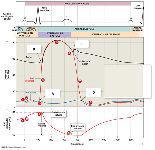
B. The first heart sound “lub” is associated with which labelled point on the Wigger’s diagram What creates this sound (1 mark)
Question 13: Cardiovascular System
The below pictures illustrate phases of haemostasis. Choose ONE of the pictures. State which phase of haemostasis is depicted. You must also provide a brief description of your chosen phase. Highlight your chosen row in bold. Note: it is not necessary to label parts of the diagrams, but you may do so if it will help you explain the phases. (2 marks total)
|
Picture |
Name of phase |
Description of phase |
|
|
|
|
|
|
|
|
|
|
|
|
Question 14: Nervous System
The following questions relate to the role and importance of a spinal reflex in the body.
A. Complete the table below to summarise the FIVE anatomical units of the reflex arc (column 1). Use the patellar reflex to provide an example for the blank anatomical units (column 2). (3 marks)
|
|
Anatomical unit |
Example (patellar reflex) |
|
1 |
Receptor |
|
|
2 |
|
|
|
3 |
Integrating centre |
|
|
4 |
|
|
|
5 |
|
|
B. Using the patellar reflex as an example, explain the purpose of this neural reflex. (2 marks)
Question 15: Nervous System
Name the region of the brain responsible for regulation of breathing and blood pressure. Would you expect damage in this region to be fatal Why (1 mark)
Question 16: Nervous System
Using the diagram provided as a prompt, outline the four steps involved in the transmission of an electrical impulse from the presynaptic neurone to the post synaptic neurone.
|
Step |
Outline |
|
1 |
|
|
2 |
|
|
3 |
|
|
4 |
|
Question 17: Urinary System
List two (2) differences between the external and internal urethral sphincters. The differences must relate to structure, function or innervation of these sphincters. (1 mark/difference; 2 marks total)
Question 18: Urinary System
A. The following table describes the three elementary steps of urine formation. Complete the missing parts of the table. (0.5 marks/cell; 3 marks total)
|
Step in urine formation |
Where does this step take place |
What happens |
|
Filtration |
|
|
|
|
|
Water and/or solutes are transported from the tubular lumen to peritubular capillaries (blood) |
|
|
Along the length of the tubular network; the exact location depends on the substance being transported |
|
Along the length of the tubular network; the exact location depends on the substance being transported B. Glucose and protein (e.g. albumin) are substances normally found in blood. Choose ONE of these molecules and complete the relevant column in the table below. Assume you are discussing what happens in a healthy person. (1 mark per box; 3 marks total)
a) Describe what happens to your selected substance at the glomerulus. Explain why/why not the substance will move into the tubule system.
b) If the substance moves into the tubule system, describe what happens to the substance along the tubule system. Explain why.
c) Do you expect to find this substance in the urine Explain why or why not.
Answer
Answer 1: Immune System
Which type of immunity is expressed in the following scenarios Choose between:
• Non-specific (first line defence)
• Non-specific (second line defence)
• Specific (antibody-mediated)
• Specific (cell-mediated immunity)
(0.5 mark each – total 1.5 marks)
|
Scenario |
Type of immunity |
|
Coughing or sneezing. |
Non-specific (first line defence)
|
|
The hypothalamus raises the body temperature above the normal range, producing fever. |
Specific (cell-mediated immunity)
|
|
Plasma cells respond to chicken pox virus |
Specific (antibody-mediated)
|
Answer 2: Immune System (2 Marks)
Choose TWO of the below scenarios and highlight these rows bold. State which general property of adaptive immunity the scenario illustrates in the second column. Select ONE phrase (from the list below) that BEST describes what is happening at a cellular level to result in this property of adaptive immunity. Write the letter corresponding to this phrase in the third column. An example is included. (0.5 mark each – total 2 marks)
|
Scenario |
General property of immunity |
Explanation |
|
Even though your body contains a relatively small number of lymphocytes, your immune system can mount a response against almost any antigen it encounters. |
Versatility |
A |
|
Generally, after an initial infection, subsequent immune responses to that antigen are faster, stronger and more sustained. |
Memory |
C |
|
You can suffer from ‘the flu’ multiple times throughout your lifetime. |
|
|
|
Your immune system can distinguish between antigens on your own cells, and those of an invading pathogen. |
Specificity |
B |
Possible phrases are listed below. Not all phrases will need to be used and only ONE phrase is required per row. If you record more than one, you will not be awarded any marks for your explanation.
A There are millions of different lymphocyte populations, each of which is sensitive to a different antigen. Lymphocytes proliferate when activated by their specific antigen.
B Each T or B cell has receptors that respond to only one antigen and ignores all others.
C Activated lymphocytes produce two groups of cells: one group that attacks the invader immediately, and another that remains inactive unless it is exposed to the same antigen at a later date.
D Each T and B cell can recognise many antigens and respond to a wide variety of possible threats.
E T cells are versatile because they produce copious quantities of antibodies that can respond to a wide variety of threats.
F Phagocytes have a reduced ability to destroy pathogens during subsequent infections.
G The immune response ignores self-antigens and targets non-self-antigens.
H B cells differentiate into T cells to create a long-lived immune response.
Answer 3: Bones
Describe the role of osteoblasts and osteoclasts in the regulation of blood calcium ion concentration. You must include the role of the relevant hormones for full marks. (3 marks)
The mechanism by which the blood calcium level is maintained by the body is known as calcium homeostasis.
The process is facilitated by the specific bone cell known as osteoblast and osteoclast and specific hormone. The osteoblast has the distinct role of absorbing calcium to make new bone matrix whereas, osteoclast is having the opposite effect as they function to secrete the stored calcium level inthe systemic circulation. The activities of these bone cells are measured by the three hormones, which includes the parathyroid hormone, calcitriol and Calcitonin. The parathyroid gland produces a parathyroid hormone in retort to the reduced level of calcium in the blood. It activates the osteoclast cells that causes reabsorption of calcium by releasing it into the blood. PTH also function by inhibiting the activity of the osteoblast that lowers the deposition of calcium into bone. The hormone directly affects the kidney to upsurge the reabsorption of calcium by triggering calcitriol. It acts on the intestine to augment the captivation of the dietary calcium. When the blood calcium is raised to the high level, the secretion of the PTH hormone is stopped. Thus, it can be said that in the condition of the overproduction of PTH, there is the release of an excessive amount of calcium into the blood. Therefore, another hormone recognized as calcitonin released by the thyroid gland has the opposite effect than PTH. It functions by inhibiting the activity of osteoclast and activating osteoblast. It also stimulates the kidney to excrete calcium. Consequently, the level of blood calcium decreases and calcium is added to the bone. Thus, the level of calcium ion in the body is regulated.
Answer 4: Muscles
Define atrophy and use an example to explain how this term relates to skeletal muscle tissue.
The term atrophy is defined as the physiological process where there is the partial or complete loss of body tissue and organ through reabsorption or breakdown of the cells, involving apoptosis. Atrophy is referred to as the reduction in the size of the cells, organ, or tissue.
In skeletal muscle tissue, due to the genetic factors, malnutrition, side effect of medication, and accident or injury, negatively impact the musculoskeletal or nervous system. Consequently, there is weakening or loss of the muscle cell, loss of fats in muscles and damage of integrity of the muscular tissue, due to which disability or immobility is prevalent. Disuse also causes rapid muscle atrophy that may occur due to prolonged illness in which immobilization of limbs is a prerequisite. Thus, it can be said that skeletal muscle atrophy is featured by the shortening of the fibre of muscle cells and damage of muscle mass.
Answer 5: Endocrine System
Think about how the renin-angiotensin-aldosterone system acts to maintain normal blood pressure. Susan has sustained damage to her adrenal glands. Consequently, her circulating levels of aldosterone are abnormally low. What do you expect the lack of aldosterone to do to Susan’s blood pressure Explain your answer. (3 marks)
Aldosterone is the type of steroid hormone which is formed by the adrenal gland. It has a crucial function of homeostatic regulation of the blood pressure, potassium level and sodium level in blood plasma. Renin-angiotensin system is considered as the major regulator of the Aldosterone, where two hormones known as renin and angiotensin controls its secretion. It is noted that Susan had an injury that damaged her adrenal gland andcaused in a reducedamount of Aldosterone in the body. As a consequence, Susan's blood pressure will decrease. It is noted that Aldosterone function to increase sodium reabsorption from the epithelial cell of kidneys and transfer it to the blood. At the same, it also makes the potassium ion travel into the kidney for its excretion in urine. Hence, when the Aldosterone level is low, it will lower the sodium level in blood due to which blood volume and blood pressure will decrease. It is articulated from the fact that Aldosteronebeing a mineralocorticoid acts by binding with its receptor present on the renal distal nephron and other sites like colon as well. Upon binding, it upsurges the absorption of sodium from the kidney mainly from the luminal cells of the cortical collecting tubules and the distal convoluted tubule, which is regarded as the major site of renal sodium reabsorption. Consequently, the high sodium level in the blood causes high blood volume and thereby blood pressure.
Answer6: Endocrine System And Urinary System
Tasma is enjoying a night out at the pub with her friends. It is well established that alcohol inhibits the secretion of ADH.
Explain how this will affect her urine output. (3 marks)
To maintain the body volume by regulating the volume of water expelled by the kidney is the chief role of the antidiuretic hormone. It has a direct function in the water reabsorption by acting the kidney tubules and concentrating salt and waste material which is excreted in the form of urine. It is articulated from the fact that discharge of antidiuretic hormone is repressed by the consumption of alcohol which in turn grounds an upsurge in urine output. Hence, Tasma is reported to be consuming alcohol in the party and it will have a direct negative effect on her urine output. Excretion of urine is noted after 20 minutes of consumption of alcohol that could result in loss of body fluid. Hence, augmentation of the urine flow from the body with continuing consumption of alcohol leads to inhibit the secretion of the antidiuretic hormone that functions in promoting concentrated urine. In the deficiency of the antidiuretic hormone, the binding with the principle cell of kidney sited in late distal tubules and collecting duct is not facilitated which causes dephosphorylation of aquaporin-2. Consequently, the movement of water in the cell is not conducted due to disturbance in the osmotic gradient thus reabsorption of water into the body is prevented. Under such circumstances, the formation of urine has diluted that result in the rise inconcentration ofions in the blood. This upsurge causes the release of antidiuretic hormone, however, owing to the high absorption of alcohol in the blood disrupt the regulatory response through overpowering the secretion of antidiuretic hormone into the blood. Thus, cause an increase in the output of urine.
Answer 7: Genetics
Duchenne muscular dystrophy (DMD) is recessive sex (or X)-linked disorder, which causes muscle degeneration and premature death. A couple is pregnant with a boy and neither has DMD. The father’s family has no history of DMD, but the mother is unsure of her genetic family history as she was adopted. The mother decides to take advantage of some of the new genomic screening tests and learns information about a large number of her genes. She finds that she is a carrier of the DMD-affected allele. (3.5 marks total)
A. What is the mother’s genotype Explain your reasoning. (1 mark)
B. What is the father’s genotype Explain your reasoning. (1 mark)
C. Explain why DMD is more common in males than females (1.5 marks)
A. For the female to be reported with the disease, it is essential to have a recessive mutated gene for DMD on both of the X chromosomes. If the genetic change is present on one of the copies of the X chromosome, she is known as a carrier. In the case, of genomic screening test mother was found to be a carrier which reflects that one of the X chromosomes carries the recessive mutated gene for DMD which has been received from either her carrier/affected mother or affected father. Hence, the genotype of the mother will be XXXx. x= mutated gene for DMD, X= normal gene for DMD.
B. It is noted that the father is not having DMD and also there is no family history regarding the prevalence of DMD. It suggests that his X chromosome does not have any recessive mutated for DMD hence is genotype will be XX Y. X= normal gene for DMD. Since the Y chromosome does not carry the gene for DMD it is normal.
C. Duchenne muscular dystrophy (DMD) is an X linked recessive disorder, where the mutated gene for causing DMD is carried on the X chromosome. Males are having one X chromosome, hence if he receives the mutated X gene from mother or father, he will have DMD. DMD is most common in males than females because as discussed DMD is X linked recessive disease that indicates that a mutated gene for DMD can only be carried by the X chromosome. Males are having only one X chromosome and a single recessive gene present on it will cause the disease. While the female can get DMD when the recessive gene is received from each of the parents as they have two X chromosomes. Hence, the probability of having DMD is common in males than females.
Answer 8: Respiratory
Describe the changes in volume and pressure inside the chest cavity during a normal, quiet exhalation.
C. Explain why these changes are occurring, and the effect upon airflow. (4 marks)
D. Is quiet exhalation an active process Briefly explain your answer. (1 mark)
A. The process of expiration is reliant on the pressure difference in lungs and the atmosphere. Three types of pressure are acted on the function of respiration, first, the atmospheric pressure which is total force exerted by the gas on the surface surrounded by air (760mmHg) second intra-alveolar pressure which total pressure of the air inside alveoli and third is intrapleural pressure which is present inside the pleural cavity of lungs. Such pressure gets changes in response to the different phases of the breathing. It is noted that during the breathing cycle, intrapleural pressure remains lower or negative to the intra-alveolar pressure however it tends to change during the expiration and inspiration with the response to the change in volume inside the chest. Exhalation is the process in which the air inside the lung cavity is expelled out through the involvement of basically two groups of muscle known as the diaphragm and the external intercostal muscles.At the time of the quiet exhalation, the pliability of lung tissue is responsible for the recoiling of the lungs. Following inspiration, diaphragm and the intercostal muscle relaxes with the response to normalization of the capacity inside the chest cavity. With the relaxation of the diaphragm and intercostal muscle, it pulls up and moves posteriorly away from the abdominal cavity, creating a less thoracic cavity and less space for the lungs. It makes the intercostal muscle relax that moves the ribs downward and inside that cause the rib care to constrict thereby decrease the volume of the thoracic cavity. The force of pleural fluid gets lowered and the contraction of the thoracic cavity causes relaxation of lungs and reduction. This decreases in volume inside the chest cavity, cause an increase in the intra-alveolar pressure, thus causing higher pressure than the atmospheric pressure. Therefore, such a pressure gradient makes the airflow from the high-pressure area to low pressure area andexpel air out of the lungs.
B. No, quiet exhalation is not an active pressure, it does not energy to push the air out of the cavity of lung rather depend on the elastic shrinking of the stretched lungs with the response to relaxation of the diaphragm and intercostal muscles.
Answer 9: Respiratory System
C. Complete the below table of partial pressures in external respiration. (2 marks)
|
|
pO2 (mmHg) |
pCO2 (mmHg) |
|
Alveoli |
104 |
40 |
|
Capillary |
40 |
45 |
D. Use to the figures above to explain how differences in partial pressures drive the direction of gas movement between the blood and alveoli. (2 marks)
In external respiration, the variance in the partial pressure of oxygen and carbon dioxide in alveoli and blood capillary is the major reason for gas exchange. It is known that despite low solubility of blood, there is a huge transformation in its partial pressure between capillary and alveolithat estimate to be approx. 64 mm Hg. In alveoli, the partial pressure is 104mm Hg while in the blood capillary is near 40 mm Hg. Such high change creates a pressure gradient that makes the oxygen move into the blood from alveoli through the respiratory membrane. The difference in the partial pressure of carbon dioxide is less than the oxygen, which estimates to be 5mm Hg. However, carbon dioxide is having a 20-fold higher solubility than oxygen in alveoli and blood. Consequently, carbon dioxide easily travels from the blood into the alveoli and the relative concentration of the gases gets equalize.
Answer 10: Cardiovascular System
A person with type B blood has been involved in a car accident and excessive bleeding necessitates a blood transfusion. Due to an error by a careless laboratory technician, the person is given type A blood. Explain what will happen. (4 marks)
For blood transfusion, the compatibility of the blood that is donated with the patient must be maintained. It is known that the blood that is donated must lack ABO and Rh D antigen that is absent in the patient. For instance, if patient of B blood group that lacks A antigen can receive blood from the person of B blood group or O blood group. However, as shown in the case where the injured person of type B blood group has received type A blood group, a chance of immune-mediated transfusion reaction may occur that arise from the immune system of the patient. In such a reaction, the incompatible RBC gets destructed which is also identified as haemolytic transfusion reaction. It can be acute that occurs immediately or may take a few days. However, it is also known that in transfusion reaction, not only RBC gets destroyed, the WBC and platelet also get destructed which is recognized as febrile non-haemolytic transfusion reaction and post-transfusion purpura respectively.
It is articulated from the fact that the individual with Type B blood group will have prepared antibodies contrary to the blood antigen which are absent in them. So, when Type A blood will be transfused in the patient, it will be sensed as an antigen by the body and antibodies against A antigen will attack the blood cells by activation of the complement system and cause Intravascular haemolysis. The major reason is the presentation of the blood group antigen on the surface of RBC. Moreover, it is also possible that Type A RBC is removed from the circulation of blood through the action of macrophage. Such RBC remained coated by the antibodies of the patient but fails to cause any immediate Intravascular haemolysis. Despite, they are identified by the IgG-Fc receptors present in macrophage and cause phagocytosis of the cell. Such reaction manifests fever, chilling, difficulty in breathing, muscle ache, nausea, chest pain and jaundice.
Answer 11: Cardiovascular (3 Marks)
To maintain adequate circulation, the average resting heart rate of a healthy non-athletic person is between 60-90 beats/minute. However, a trained athlete’s heart rate is usually about 40 beats/min (sometimes as low as 30 bpm!). Despite this, adequate circulation is maintained in these athletes. Using the terms cardiac output and stroke volume, explain how this is possible. (3 marks)
To maintain the circulation of blood, it is crucial to have an adequate cardiac output which is the volume of blood being impelled by the ventricle in one minute. It is measured by multiplication of stroke volume and heart rate. In a normal person, the heart rate is 60-90 beats/minutes. However, in an athlete, the heart rate is only <40 bpm at rest to >200 bpm. The major reason is the prolonged training that increases the stroke volume and cardiac output by 10-fold than the normal person. There is the enlargement of the cardiac chamber due to the excessive exercise which is being regarded as the adaptability for normal blood circulation. Therefore, as a result, the stroke volume increase as the consequence of upsurge in the ventricular end-diastolic volume and sympathetically mediated decrease in end-systolic volume. The increase in the contraction of the ventricle, the stroke volume increase from 70 to 130 mL and cardiac output augments to 19.5L/min that is 5-6 times higher than the resting rate. Hence, despite the low heart rate in athletes, the high cardiac output and stroke volume maintain the flow of blood into the heart and tissue. The heart becomes more efficient as it is capable of pumping a larger volume of blood with each contraction of the ventricle. Additionally, with long term training, the demand for myocardial oxygen decreases through which amount of blood pumped to sufficient to maintain the blood circulation.
Answer 12: Cardiovascular
C. The Wigger’s diagram (below) represents pressure and volume relationships in the left side of the heart during one cardiac cycle. To answer this question, you should focus on the changes in pressure in different chambers of, or vessels leading from, the left side of the heart.
For TWO of the labelled points, state which valve is open/closed at that time. Highlight your chosen point in bold. You must also explain WHY that valve opened/closed and where the blood is flowing immediately after this point in time, due to the change in position of the valve. An example is provided. (4 marks)
|
Label |
Valve (name and open/closed) |
Explanation |
|
A |
Left AV/bicuspid close |
When the final period of diastole of the atrium is over, it gets contracted due to the rise of pressure and blood drifts into the ventricles that cause a slight increase in its pressure. It serves as the priming mechanism to increase the ventricular preload before ejection. Therefore, when ventricular pressure becomes high than the atrium, the AV valve is forced to get closed in order to avoid the backflow of the heart. |
|
B |
Aortic value opens |
When the ventricular pressure surpasses the pressure present in the aorta, that is at the time of 75-80mmHg ventricular pressure, it exceeds the aortic pressure causing the opening of the aortic valve that forces the blood to travel to aorta ejected from the ventricle. |
|
C |
|
|
|
D |
Left AV/bicuspid valve opens |
The pressure in the left ventricle becomes lower than that in the left atrium. Blood flows from the left atrium to the left ventricle. |
Answer 13: Cardiovascular System
The below pictures illustrate phases of haemostasis. Choose ONE of the pictures. State which phase of haemostasis is depicted. You must also provide a brief description of your chosen phase. Highlight your chosen row in bold. Note: it is not necessary to label parts of the diagrams, but you may do so if it will help you explain the phases. (2 marks total)
|
Label |
Valve (name and open/closed) |
Explanation |
|
A |
Left AV/bicuspid close |
When the final period of diastole of the atrium is over, it gets contracted due to the rise of pressure and blood drifts into the ventricles that cause a slight increase in its pressure. It serves as the priming mechanism to increase the ventricular preload before ejection. Therefore, when ventricular pressure becomes high than the atrium, the AV valve is forced to get closed in order to avoid the backflow of the heart. |
|
B |
Aortic value opens |
When the ventricular pressure surpasses the pressure present in the aorta, that is at the time of 75-80mmHg ventricular pressure, it exceeds the aortic pressure causing the opening of the aortic valve that forces the blood to travel to aorta ejected from the ventricle. |
|
C |
|
|
|
D |
Left AV/bicuspid valve opens |
The pressure in the left ventricle becomes lower than that in the left atrium. Blood flows from the left atrium to the left ventricle. |
Answer 14: Nervous System
The following questions relate to the role and importance of a spinal reflex in the body.
C. Complete the table below to summarise the FIVE anatomical units of the reflex arc (column 1). Use the patellar reflex to provide an example for the blank anatomical units (column 2).
(3 marks)
|
|
Anatomical unit |
Example (patellar reflex) |
|
1 |
Receptor |
Muscle spindle |
|
2 |
Sensory (afferent neuron) |
|
|
3 |
Integrating centre |
Spinal cord |
|
4 |
Motor efferent neurone |
|
|
5 |
Effector |
Extrafusal cord |
D. Using the patellar reflex as an example, explain the purpose of this neural reflex. (2 marks)
A patellar reflex takes place through the transmission of the signal arise due to the stretch of the quadricep muscle. Initially, stretch take place, it is sensed by the spindle muscle and cause activation of sensory neuron and signal is travelled from at the level of spinal cord to the motor neuron. Such signal is known to regulate the quadricep contraction and reduced the movement of leg. Therefore, patellar reflex is regarded as the monosynaptic reflex due to the direct association of sensory neuron and motor neuron. Therefore, the main purpose of the neural reflex is to carry the sensory information to the spinal cord from the receptor and then to carry it to the effector organ from the spinal cord thus creating the reflex arc.
Answer 15: Nervous System
Name the region of the brain responsible for regulation of breathing and blood pressure. Would you expect damage in this region to be fatal Why (1 mark)
The medulla oblongata is accountableto control breathing and blood pressure. Yes, injury to this area of the brain is expected to be fatal because it will block the signal that is being transferred to and from the brain and spinal cord for the control of vital body functions like breathing and heart functioning. Moreover, Medulla oblongata is considered to contain cardiac,vasomotor and respiratory centres that control the heart rate, rate of breathing and other hence injury to medulla oblongata could cause loss of life-sustaining body function.
Answer 16: Nervous System
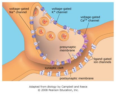
Using the diagram provided as a prompt, outline the four steps involved in the transmission of an electrical impulse from the presynaptic neurone to the post synaptic neurone. (4 marks)
|
Step |
Outline |
|
1 |
Arrival of action potentialThe first step in the transmission of the signal from presynaptic to postsynaptic neurons is the arrival action potential in the presynaptic neuron. When the excitatory signal is sensed by the neuron, it tends to activate the voltage-gated channel for sodium-ion to open. At the resting stage, inside the concentration of sodium is low so it will flow inside the cells and making the membrane potential more positive and thus giving rise to the action potential. This, in turn, activate the K+ ion channel and it diffuses out of the axon and causes closing the sodium channel to get inactivated at that point however the first action potential causes a large electrical change that reaching to the threshold is not possible and triggers the opening of the sodium channel. This causes downswing of the action potential at the synaptic end. |
|
2 |
The fusion of the vesicle with the plasma membraneThere are two categories of the neurotransmitter the neuropeptide and smaller amino acids which are produced in the cell body of the neuron as it requires peptide bond formation and presynaptic terminal respectively. These molecules are stored in the form of the vesicle in the terminal of axon until the arrival of action potential for their release. In response to the occurrence of action potential, neurotransmitter gets fused with the plasma membrane. |
|
3 |
Release of neurotransmitter into the synaptic cleft When the action potential depolarizes the nerve, impulse tends to opens the voltage-gated calcium channels thatexist in the synaptic end bulb. As calcium is more intense at the extracellular fluid, effluxes to the open channels, this increases the amount inside the presynaptic neuron and trigger signal for exocytosis of the synaptic vesicle. Increasedconcentration of ca+ present in the terminal neuron triggers the fusion of vesicle and plasma membrane that result in the releases of neurotransmitters inthe synaptic cleft. |
|
4 |
Binding of neurotransmitter to receptorIn the fourth step, the neurotransmitter molecule gets diffusedin the synaptic cleft and associates with the neurotransmitter receptor in the plasma membrane of postsynaptic neuron. With binding of the neurotransmitter molecule to the receptor on ligand-gated channels causes its opening and this makes potassium and sodium ion to flow in the membrane. This, in turn, causes a change in voltage of membrane which is known as postsynaptic potential, where neurotransmitters are released from the receptor thus closes the channels and triggers the final stage of synaptic transmission with the efflux of neurotransmitters out of the synaptic cleft. This is for the taken up by the synaptic terminal or degraded by the enzyme. |
Answer17: Urinary System<
List two (2) differences between the external and internal urethral sphincters. The differences must relate to structure, function or innervation of these sphincters. (1 mark/difference; 2 marks total)
|
Factor |
External sphincter |
Internal sphincter |
|
Structure |
It is made up of skeletal muscle tissue and located below the bladder surrounding the urethra. |
It is made up of smooth muscle and surrounds the neck of the bladder. |
|
Function |
It functions to control urination involuntarily. |
It is voluntary and functions to open the bladder to the urethra and relaxesthat let pass the urine. |
Answer 18: Urinary System (6 Marks)
C. The following table describes the three elementary steps of urine formation. Complete the missing parts of the table. (0.5 marks/cell; 3 marks total)
|
Step in urine formation |
Where does this step take place |
What happens |
|
Filtration |
In the glomerulus and then to the bowmen capsule. |
Blood gets filtered in the glomerulus where substance like water and waste material (nitrogenous) easily move inside of the glomerulus and enter bowmen capsule while other substance like cell and serum albumin gets filtered. |
|
Reabsorption |
The proximal/distal convoluted tubules, loop of Henle, the collecting duct. |
Water and/or solutes are transported from the tubular lumen to peritubular capillaries (blood) |
|
Secretion |
Along the length of the tubular network; the exact location depends on the substance being transported |
The solutes like hydrogen ion and creatinine are tended to eliminate from the blood through the passage from the peritubular capillary and enters the collecting duct. |
D. Glucose and protein (e.g. albumin) are substances normally found in blood. Choose ONE of these molecules and complete the relevant column in the table below. Assume you are discussing what happens in a healthy person. (1 mark per box; 3 marks total)
d) Describe what happens to your selected substance at the glomerulus. Explain why/why not the substance will move into the tubule system.
e) If the substance moves into the tubule system, describe what happens to the substance along the tubule system. Explain why.
f) Do you expect to find this substance in the urine Explain why or why not.
|
|
Glucose |
Protein |
|
a) Glomerulus
|
In the glomerulus, the blood is known to flow through it and with the action of blood pressure, the glucose molecule is transferred to the wall of the capillary and enters the Bowman's capsule. It is noted that such an initial step helps in the removal of the waste products present in the blood and simultaneously aid in preventing loss of cells like RBC and protein. However, along with that it also removes glucose present in the bloodstream. |
|
|
b) Tubule system
|
It is known that the tubular portion of the nephron comprises proximal tubules, the loop of Henle and distal tubules. Among those, the distal tubules and proximal tubules have the opposite function where the distal tubules are known to secrete the waste solutes into the urine while the proximal tubules have a major role for reabsorbing solutes into the blood. Hence, the reabsorption of the glucose molecules occurs in the proximal tubules which are directly linked with the Bowman's capsule. The cell lining of the proximal tubules tends to reabsorb glucose through the apical membrane or shunter transporter. Sodium dependent glucose cotransporters are found embedded in the apical membrane of the proximal tubules that function as a pump to drive the sodium ion efflux and potassium influx and creates a concentration gradient of the ions. This gradient causes sodium-dependent glucose cotransporter 2 to transport glucose molecules into the cell. And once it is inside the cell, GLUT2 help in returning to the bloodstream. |
|
|
c) Urine
|
In urine, the glucose molecule is not found and it only contains water, waste material and urea. Glucose gets filtered through the glomerulus and found in the glomerulus filtrate which further gets reabsorbed by the bloodstream. Through the action of GLUT 2, the concentrated glucose molecules on the cell of PCT is transferred back to the blood flow and does not enter the urine.
|
|